Chapter 2. Basic Cardiac
Chapter Contributors
- James Hwang, MD, RDMS, RDCS, FACEP
- Jonathan Wallace, MD, RDMS
- Rayna Sharma, BScKin
- Adam Guthrie, BKin, MMgmt
Background
Indications for Bedside Echo
- Cardiac Arrest
- Unexplained Hypotension
- Syncope
- Dyspnea
- Chest Pain
- Trauma
Scope of Basic Cardiac Bedside Ultrasound
Movie 2.1
Normal Subxiphoid View
Basic echocardiography is a core application of bedside ultrasound and a key element of sonography algorithms, such as the EFAST exam (Chapter 1. FAST) and the RUSH protocol (Chapter 5. RUSH).[1][2][3] Although comprehending image orientation[4] and acquiring images can be somewhat challenging, clinician performed bedside echocardiography has a major impact on our ability to detect cardiac abnormalities and on patient care, and can be life-saving.[5][6] Bedside echocardiography provides clinicians with time-sensitive anatomic and physiologic information in a variety of cardiac-related scenarios, including cardiac arrest, unexplained hypotension, syncope, shortness of breath, and chest pain[6:1][7][8][9][10][11] #Movie 2.2. Bedside echocardiography can help clinicians risk-stratify patients and further guide resuscitative efforts.[12][13] At times, the information and disorders noted on bedside cardiac ultrasound are vast and may surpass what performing clinicians are able to appreciate and integrate. As per the American Society of Echocardiography (ASE) – American College of Emergency Physicians (ACEP) Consensus Statement, the ability to assess global left ventricular function, to detect pericardial effusion, and to assess for right heart dilatation (chamber sizes) are within the scope of clinicians and can help answer critical patient management questions.[8:1]
Movie 2.2
Severely Depressed EF
Technique
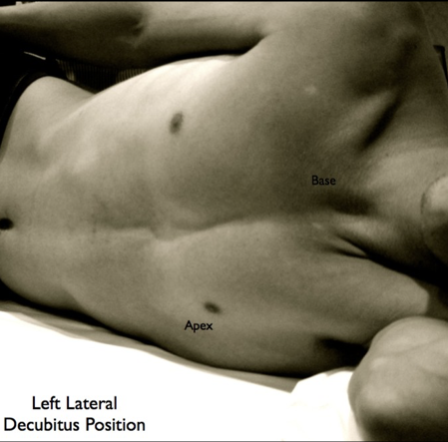
Left lateral decubitus position (#Image 2.2) will improve view in parasternal and apical views.
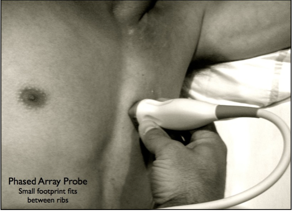
The heart lies obliquely in the chest with the apex pointing towards the left hip and the base of the heart oriented towards the right shoulder (#Figure 2.1). The heart is imaged from multiple different views and the findings seen on one view should be confirmed or refuted with additional views. A 2 to 5 MHz phased array probe is used when attempting to obtain acoustic windows of the heart, as the smaller footprint (flat square face) more easily fits in between the ribs (#Image 2.1).
Figure 2.1
Heart
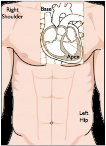
Image 2.1
Parasternal Long Axis View
There are four basic cardiac views: parasternal long axis, parasternal short axis, apical 4-chamber, and subcostal. If possible, the patient should be rolled into the left lateral decubitus position, as this brings the heart closer to the anterior chest wall and improves imaging (#Image 2.2).
Image 2.2
Left Lateral Decubitus Position
Parasternal Long Axis (PSLA)
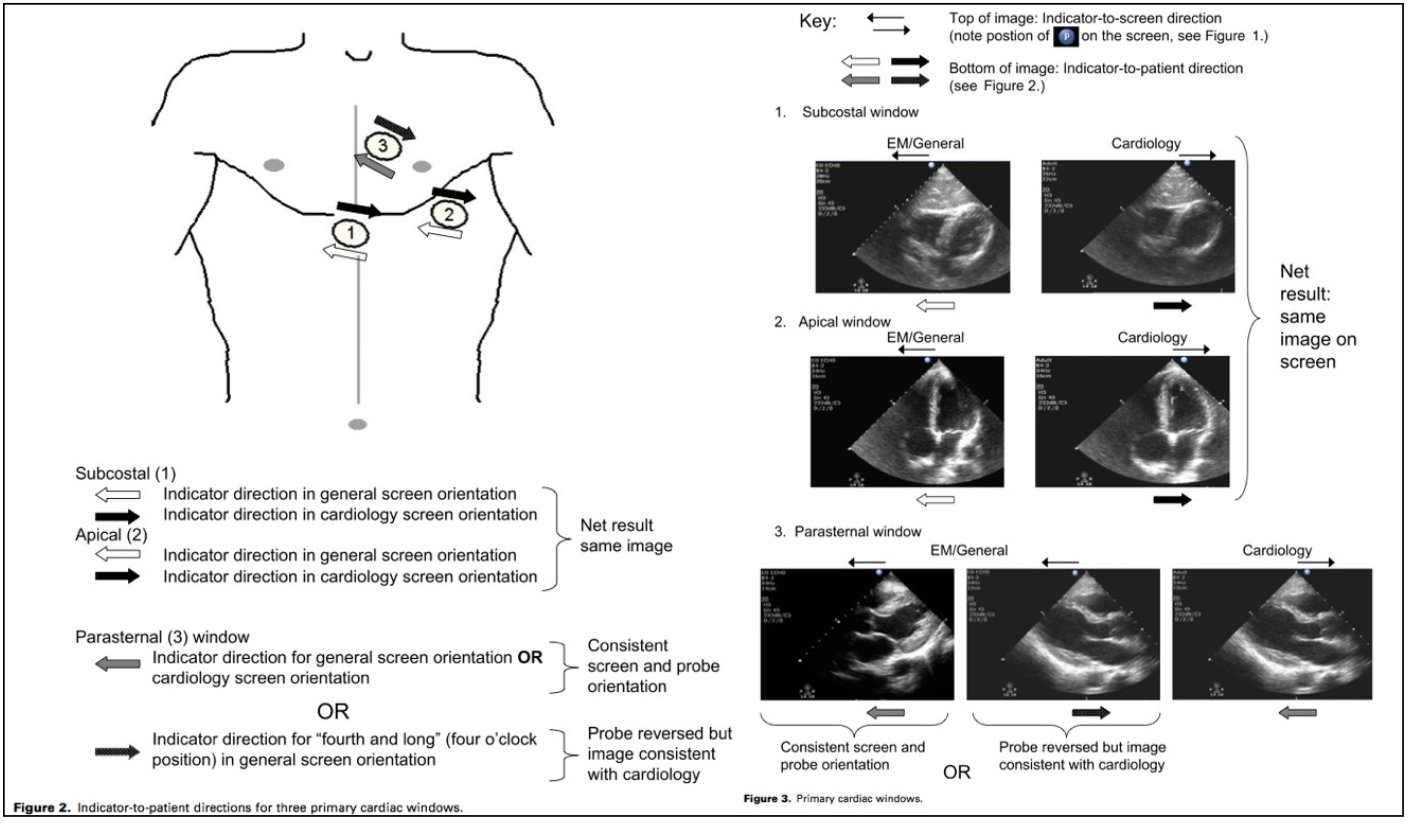
The parasternal long axis view is obtained by placing the probe to the left of the sternum with the probe marker pointing towards the patient’s right shoulder. The probe is then gently dragged over the chest wall from the 2nd to 5th intercostal spaces, searching for the best acoustic window (#Movie 2.3). The PSLA view is consistently obtainable and provides an excellent view of the left atrium (LA), left ventricle (LV), and aortic outflow tract.
Movie 2.3
PSLA View
Parasternal Short Axis (PSS)
Once the parasternal long axis view is acquired, the probe can then be rotated clockwise 90° with the probe marker pointing towards the patient’s left shoulder to obtain the parasternal short axis (PSSA) view (#Movie 2.4). The PSSA view is an ideal view for assessing global LV function.
Movie 2.4
PSSA View
Apical 4-Chamber (A4C)
The apical 4-chamber (A4C) view is obtained by placing the probe inferior and lateral to the left nipple in men or under the left breast in women. The probe marker is pointed towards the patient’s left axilla and the face of the transducer is angled up towards the base of the heart. (#Movie 2.5).
- The probe represents the entry point, and you must align it such that tip of the spear (sound waves) will exit through the right shoulder (not through the spine or scapula)
- This trick helps you visualize correct alignment of your probe for the long axis of the heart to acquire the A4C view.
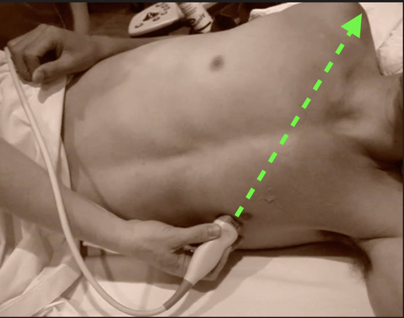
The A4C view is often the most difficult view to acquire but offers valuable information about all four chambers and their relative sizes.
The easiest means of dealing with both breast tissue and most bras is to ask the patient to retract the left breast for you. This approach offers multiple benefits:
- it maximizes comfort and dignity for the patient
- it removes the burden and distraction of retracting breast tissue yourself
- it exposes the inframammary fold which is an ideal landmark, frequently correlating to the ideal rib space for the A4C window.
Ultrasound waves generally do not penetrate clothing, so if the patient is unable to adequately retract material to expose the window you are seeking, the clothing will need to be removed.
Movie 2.5
Apical 4-Chamber View
**CORRECTION** to video commentary: *The probe marker is pointed towards the __patient’s left axilla__...* >[!note]- Additional Details Most modern PoCUS machines now use the *cardiology convention* when selecting the cardiac probe (phased array). In cardiology convention the ultrasound image is flipped horizontally. You can confirm the image orientation by noting which side of the image the manufacturer's logo / probe marker is on. Marker icon on left is conventional for PoCUS scanning for non-cardiac applications (and historically the emergency-medicine convention during cardiac scanning.) Marker icon on right (mirror image) is the cardiology convention which dominates in advanced to fellowship level cardiac PoCUS. Hence the discrepancy in the narration of movie 2.5Subcostal (Sx)
The subxiphoid view is obtained by placing the probe just below the xiphoid process with the probe marker pointing towards the patient’s left hip. The subxiphoid view uses the left lobe of the liver as an acoustic window and involves "flattening" the probe such that the sound waves travel just below the anterior chest wall. Placing the hand over the probe, as opposed to holding the probe like a pencil, as in other views, allows for the requisite angling of the probe where it is almost flat against the abdominal wall (#Movie 2.6).
Movie 2.6
Subxiphoid View
For a more comprehensive discussion reviewing cardiac ultrasound image orientation, see Moore’s Special Contribution: Current Issues with Emergency Cardiac Ultrasound Probe and Image Conventions[4:1] (#Image 2.3).
Image 2.3
Cardiac Window
Left Ventricular Function
Global left ventricular function can be accurately assessed by PoCUS. Scenarios where LVF may be useful:
- Chest pain
- Dyspnea
- Unexplained hypotension
- Cardiac arrest
Assessment of global left ventricular function is a fundamental application of basic bedside echocardiography and helps predict clinical outcomes for a variety of disease states. The ability to assess a patient’s overall left ventricular function — from cardiac standstill (#Movie 2.7) to a hyperdynamic ejection fraction (#Movie 2.8) — allows clinicians to better manage patients who present with chest pain, dyspnea, unexplained hypotension or cardiac arrest.[14][15]
Movie 2.7
Cardiac Standstill
- Notice a possible subtle motion in the tricuspid leaflets at about the 8-10 cm depth level. Subtle motion like this is common, but in the absence of lack of myocardial motion this is still classified as cardiac standstill.Movie 2.8
Hyperdynamic EF
While regional wall motion abnormalities and quantitative measurements may be beyond the scope of some clinicians, the visual (qualitative) estimate of global left ventricular systolic function is not.[8:2][16]
Assessment of systolic function is derived from the visual assessment of endocardial border excursion and myocardial thickening, as seen from multiple views (#Movie 2.9, #Movie 2.10, #Movie 2.11 and #Movie 2.12).
Movie 2.9
Parasternal Long
Movie 2.10
Parasternal Short
Movie 2.11
Apical 4
Movie 2.12
Subxiphoid 4
Classification of LV function can be simplified into the following by Ejection Fraction (EF)
- severely depressed (EF < 30%), (#Movie 2.13, #Movie 2.14)
- mild-moderately depressed (EF = 30-55%), (#Movie 2.15)
- and normal (EF > 55%)[15:1] (#Movie 2.16)
Movie 2.13
Severely Depressed EF
Movie 2.14
Severely Depressed EF
Movie 2.15
Moderately Depressed EF
Movie 2.16
Normal EF
Ejection fractions in excess of 70% are considered hyperdynamic, which may appear as near obliteration of the ventricular cavity during systole (#Movie 2.17).
Movie 2.17
Hyperdynamic with Increased EF
Do not confuse with tachycardia and normal EF. (LV walls appear to be touching: hyperdynamic)
The finding of a hyperdynamic heart may suggest hypovolemia or vasodilation and should prompt the clinician to initiate volume resuscitation. It is important, however, to differentiate a hyperdynamic heart from one that is tachycardic but with a normal EF% (#Movie 2.18 and #Movie 2.19). With appropriate education and training, clinicians can differentiate between normal and severely depressed left ventricular systolic function similar to cardiologists.[15:2] Moore et al demonstrated that with focused training, emergency physicians can accurately determine left ventricular function in hypotensive patients.[15:3]
Movie 2.18
Tachycardia with Normal EF
Do not confuse with hyperdynamic with increased EF (LV does not collapse: tachycardia)
Movie 2.19
Tachycardia with Normal EF
Do not confuse with hyperdynamic with increased EF (LV does not collapse: tachycardia)
Although this study showed good agreement between emergency physicians and cardiologists for patients with normal and severely depressed left ventricular function, emergency physicians had more trouble categorizing patients with moderately depressed LV function (#Movie 2.20). This validates the ability of clinicians to identify extremes of LV dysfunction, but underscores the need for clinicians to recognize their limitations and to obtain consultative studies when indicated.
Movie 2.20
Moderately Depressed EF
One Minute Ultrasound EPSS Demonstration
Pericardial Effusion
- Ultrasound is an ideal modality for detecting pericardial effusions.
- Tamponade is a clinical diagnosis that depends more on pressure and physiology than size of effusion.
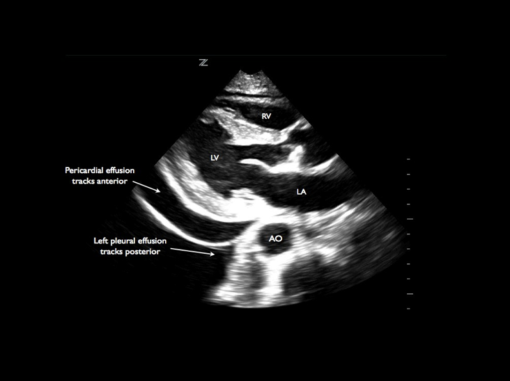
- Pericardial and pleural effusions can frequently be differentiated based on location of fluid.
Pericardial fluid typically appears as an anechoic space between the epicardium and the pericardium (#Image 2.4). Pericardial effusions are caused by a variety of disorders (infection, malignancy, connective tissue disease, renal failure, trauma) and may also develop after cardiac surgery or invasive cardiac procedures (pacemaker placement, cardiac catheterization). Ultrasound is an ideal modality to assess for the presence of pericardial fluid and its impact on right heart filling.[3:1][17][18] Pericardial effusions are not an uncommon diagnosis in patients presenting with dyspnea or hypotension.[11:1][19] Although cardiac tamponade is largely a clinical diagnosis, bedside echocardiography may demonstrate findings suggestive of impending tamponade prior to the development of physical examination findings and hemodynamic compromise.[20][21][22] The amount of fluid required to impair filling and to cause circulatory failure depends on the rate of accumulation. Pericardial effusions may be graded as:
- Small (<0.5cm) (#Movie 2.21, #Movie 2.22)
- Moderate (0.5-2.0cm) (#Movie 2.23, #Movie 2.24)
- Large (>2cm) (#Movie 2.25, #Movie 2.26)[23][24][25]
Image 2.4
Pericardial Effusion
Movie 2.21
Small Effusion
Movie 2.22
Small Effusion
Movie 2.23
Moderate Effusion
##### Movie 2.24 **Moderate Effusion**Movie 2.25
Large Effusion with RV collapsing
##### Movie 2.26 **Large Effusion** with RV collapsingWhile large effusions are often circumferential, it is important to recognize that effusions can be focal, organized, or loculated (#Movie 2.27 and #Movie 2.28).
Movie 2.27
Anterior Effusion
Movie 2.28
Anterior Effusion
A potential pitfall is differentiating between epicardial fat pads and true pericardial effusions. Epicardial fat pads are:
- often visualized anteriorly
- mostly hypoechoic in appearance but often have some echogenicity (#Movie 2.29).
Movie 2.29
Epicardial Fat Pad
Numerous studies have demonstrated that emergency physician–performed emergency echocardiography has sensitivities approaching 100% for the detection of pericardial effusions.[12:1][19:1][26] When compared with expert over-read of images, emergency physician–performed emergency echocardiography for effusion has a sensitivity of 96% to 100%, a specificity of 98% to 100%, a positive predictive value of 93% to 100%, and a negative predictive value of 99% to 100%. The echocardiographic findings consistent with cardiac tamponade include the following:
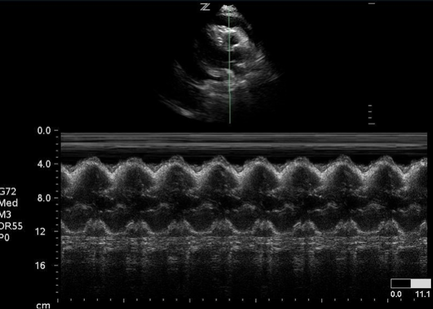
- right ventricular (RV) free wall inversion during ventricular diastole. This is the hallmark finding. (#Image 2.5, #Movie 2.30, #Movie 2.31)
- right atrial (RA) inversion during ventricular systole (more common and one of the earliest findings)
- increased respiratory variation of mitral or tricuspid inflow velocities (inspiratory decreases of greater than 25% on mitral inflow or greater than 40% on tricuspid inflow)
- a dilated inferior vena cava (IVC) with decreased respirophasic variation (#Movie 2.32).[24:1][25:1][27]
Image 2.5
RV Collapse on M-Mode - Tamponade
Movie 2.30
RV Collapse Due to Tamponade
Movie 2.31
RV Collapse
##### Movie 2.32 **Plethoric IVC in Tamponade**It is important not to confuse ventricular or atrial systole with diastolic collapse. While large pleural effusions may be misinterpreted as pericardial effusions, the descending thoracic aorta can be used to differentiate the two diagnoses.
Recall that the descending limb of the thoracic aorta is outside the pericardium, in the mediastinum. A pericardial effusion inside the pericardial sac will attempt to separate the heart and descending aorta. Whereas a pleural effusion outside the mediastinum will accumulate adjacent to the heart and descending aorta.
On the PSLA view, pleural effusions run posterior or lateral to the descending thoracic aorta, while pericardial effusions track anteriorly or medially (#Movie 2.33, 2.34).
Movie 2.33
Left Pleural and Pericardial Effusions
Movie 2.34
Left Pleural and Pericardial Effusions
Right Heart Dilation
Common causes of right heart dilatation:
- PE
- RV infarction
- Pulmonary hypertension
- COPD
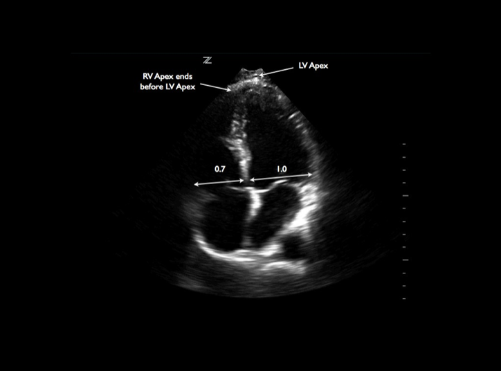
Normal RV:LV ratio = 0.6:1.0
Patients with right ventricular (RV) dysfunction can be difficult to diagnose and challenging to manage. Patients with right heart failure may worsen with aggressive fluid resuscitation, and identification and reversal of the etiology for RV dysfunction is key.[28][29] The thin-walled RV is extremely sensitive to load and, as such, small changes in pressure lead to large changes in volume. RV dilatation is the normal response to RV pressure or volume overload.[24:2] While assessing for RV systolic dysfunction or for paradoxical septal motion (#Movie 2.35) can be challenging for many clinicians, assessing for RV dilatation (chamber size) is less so.[8:3] Typically, the RV is smaller than the LV, with an RV-to-LV ratio of 0.6:1.0 (#Image 2.6). When the RV is noted to be equal in size to the LV, the RV is moderately dilated (#Movie 2.36, 2.37).
Movie 2.35
Parasternal Short with Septal Flattening
Image 2.6
Normal RV:LV Size Ratio
Movie 2.36
RV Dilation
Movie 2.38 - RV Dilation
When the RV is larger than the LV, severe RV dilatation is present[30] (#Movie 2.38, 2.39, 2.40, 2.41). The apical 4-chamber view is used to compare RV and LV sizes, and the relative sizes are compared at the tips of the atrioventricular valves in diastole. When RV dilatation is present, the RV apex is shifted closer to, or even encompasses, the LV apex.[25:2]
In the appropriate clinical setting and when combined with other variables, RV dilatation may suggest RV outflow tract obstruction due to pulmonary embolism.[31] Other causes of right ventricular dilatation must also be considered, including:
- RV infarction
- pulmonary hypertension
- chronic obstructive pulmonary disease
Historically PoCUS was not sufficiently sensitive (reported sensitivity of around 60-70%) for the detection of pulmonary embolism and thus, cannot exclude it as a diagnosis.[29:1][30:1][31:1][32]
However, in a landmark study in XXXX, it was discovered that as part of a scanning protocol and with careful application of other features, PoCUS is able to narrow the gap, identifying secondary findings such as presence of DVTs, wedge infarcts, and other clinical symptoms to better rule in or exclude PE in specific circumstances.
A chapter on Pulmonary Embolism will added later in the IBUS 2nd Edition Project.
Patients with pulmonary embolism and evidence of right-heart dysfunction, however, have increased morbidity and mortality, and bedside echocardiography can be used to risk-stratify and to better manage the hemodynamic issues in these patients.[30:2][31:2][32:1][33]
Movie 2.38
RV Dilation and Failure
Movie 2.39
RV Dilation and failure due to PE - clot in RA
Movie 2.40
RV Dilation and failure
Movie 2.41
RV pressure overload with flattening & reversal of septum
Conclusion
- Bedside echocardiography can be life-saving and dead sexy.
- It is essential to recognize one's limitations with cardiac ultrasound.
Bedside echocardiography provides clinicians with immediate structural and physiologic data that can be life-saving. [5:1][6:2] The ability to assess patients for pericardial effusions, global left ventricular function, and right heart dilatation can provide answers to critical questions, risk-stratify patients, and further guide resuscitative efforts. It is essential, however, to recognize one’s limitations when performing basic cardiac ultrasound and to obtain consultant-performed echocardiography when appropriate.
References
American College of Emergency Physicians. Emergency ultrasound guidelines – 2008. Ann Emerg Med. 2009;53:550-570. ↩︎
Hauser AM. The emerging role of echocardiography in the emer- gency department. Ann Emerg Med. 1989;18:1298-1303. ↩︎
Mayron R, Gaudio FE, Plummer D, et al. Echocardiography per- formed by emergency physicians: impact on diagnosis and therapy. Ann Emerg Med. 1988;17:150-154. ↩︎ ↩︎
Moore CL. Special Contribution: Current Issues with Emergency Cardiac Ultrasound Probe and Image Conventions. Acad Emerg Med. 2008;15(3):278-84 ↩︎ ↩︎
Kimura BJ, Bocchicchio M, Willis CL, DeMaria AN. Screening car- diac ultrasonographic examination in patients with suspected cardiac disease in the emergency department. Am Heart J. 2001;142(2):324-330. ↩︎ ↩︎
Kaul S, Stratienko AA, Pollack SJ, et al. Value of two-dimensional cardiac ultrasound for determining the basis of hemodynamic com- promise in critically ill patients: a prospective study. J Am Soc Echo- cardiogr 1994;7:598-606. ↩︎ ↩︎ ↩︎
Stahmer SA. The ASE position statement in echocardiography in the emergency department. Acad Emerg Med. 2000;7:306-308. ↩︎
Labovitz AJ, Noble VE, Bierig M, et al. Focused Cardiac Ultra- sound in the Emergent Setting: A Consensus Statement of the Ameri- can Society of Echocardiography and American College of Emer- gency Physicians. J Am Soc Echocardiogr 2010;23(12):1225-30. ↩︎ ↩︎ ↩︎ ↩︎
Rose JS, Bair AE, Mandavia D, et al. The UHP ultrasound protocol: a novel ultrasound approach to the empiric evaluation of the undiffer- entiated hypotensive patient. Am J Emerg Med. 2001;19:299-302. ↩︎
Jones AE, Tayal VS, Sullivan DM, et al. Randomized, controlled trial of immediate versus delayed goal-directed ultrasound to identify the cause of nontraumatic hypotension in emergency department pa- tients. Crit Care Med. 2004;32(8):1798-1800. ↩︎
Blaivas M. Incidence of pericardial effusion in patients presenting to the emergency department with unexplained dyspnea. Acad Emerg Med. 2001;8:1143-1146. ↩︎ ↩︎
Mandavia DP, Hoffner RJ, Mahaney K, et al. Bedside echocardi- ography by emergency physicians. Ann Emerg Med. 2001;38(4):377-382. ↩︎ ↩︎
Blaivas M, Fox J. Outcome in cardiac arrest patients found to have cardiac standstill on the bedside emergency department echo- cardiogram. Acad Emerg Med. 2001;8:616-621. ↩︎
Jones AE, Tayal VS, Kline JA. Focused training of emergency medicine residents in goal-directed echocardiography: a prospective study. Acad Emerg Med. 2003;10:1054-1058. ↩︎
Moore CL, Rose GA, Tayal VS, et al. Determination of left ven- tricular function by emergency physician echocardiography of hy- potensive patients. Acad Emerg Med. 2002;9:186-193. ↩︎ ↩︎ ↩︎ ↩︎
Randazzo MR, Snoey ER, Levitt AM, et al. Accuracy of emer- gency physician assessment of left ventricular ejection fraction and central venous pressure using echocardiography. Acad Emerg Med 2003;10(9):973-7. ↩︎
Martin RP, Rakowski H, French J, et al. Localization of pericardial effusion with wide angle phased array echocardiography. Am J Car- diol. 1978;42:904-912. ↩︎
Mazurek B, Jehle D, Martin M. Emergency department echocardi- ography in the diagnosis and therapy of cardiac tamponade. J Emerg Med. 1991;9:27-31. ↩︎
Tayal VS, Kline JA. Emergency echocardiography to detect peri- cardial effusion in patients in PEA and near-PEA states. Resuscitation.2003;59(3):315-318. ↩︎ ↩︎
Levine MJ, Lorell BH, Diver DJ, Come PC. Implications of echocar- diographically assisted diagnosis of pericardial tamponade in contemporary medical patients: detection before hemodynamic embar- rassment. J Am Coll Cardiol. 1991;17:59-65. ↩︎
Tsang TSM, Oh JK, Seward JB. Diagnosis and management of cardiac tamponade in the era of echocardiography. Clin Cardiol. 1999;22:446-452. ↩︎
Blaivas M, Graham S, Lambert MJ. Impending cardiac tampon- ade: an unseen danger? Am J Emerg Med. 2000;18:339-340. ↩︎
Shabetai R. Pericardial effusion: haemodynamic spectrum. Heart. 2004;90:255-256. ↩︎
Otto CM. Textbook of Clinical Echocardiography. 3rd ed. Phila- delphia: Elsevier– Saunders; 2004. ↩︎ ↩︎ ↩︎
ASE Committee Recommendations. Recommendations for cham- ber quantification: a report from the American Society of Echocardi- ography’s Guidelines and Standards Committee and the Chamber Quantification Writing Group, Developed in Conjunction with the European Association of Echocardiography, a Branch of the Euro- pean Society of Cardiology. J Am Soc Echocardiography. 2005;18(12):1440-1463. ↩︎ ↩︎ ↩︎
Plummer D, Brunette D, Asinger R, Ruiz E. Emergency department echocardiography improves outcome in penetrating cardiac injury. Ann Emerg Med. 1192;26:709-712. ↩︎
Gillam LD, Guyer DE, Gibson TC, et al. Hydrodynamic compres- sion of the right atrium: a new echocardiographic sign of cardiac tamponade. Circulation. 1983;68:294-301. ↩︎
Kucher N, Goldhaber SZ. Management of massive pulmonary embolism. Circulation 2005;112:e28-32. ↩︎
Torbicki A, Perrier A, Kostantinides S, et al. Guidelines on the di- agnosis and management of acute pulmonary embolism. Eur Heart J 2008;29:2276-315. ↩︎ ↩︎
Kasper W, Konstantinides S, Geibel A, et al. Prognostic signifi- cance of right ventricular afterload stress detected by echocardiogra- phy in patients with clinically suspected pulmonary embolism. Heart. 1997;77:346-349. ↩︎ ↩︎ ↩︎
Nazeyrollas P, Metz D, Jolly D, et al. Use of transthoracic echo- cardiography combined with clinical and electrocardiographic data to predict acute pulmonary embolism. Eur Heart J. 1996;17:779-786. ↩︎ ↩︎ ↩︎
Roy PM, Colombet I, Durieux P, Chatellier G, Sors H, Meyer G. Systematic review and meta-analysis of strategies for the diagnosis of suspected pulmonary embolism. Br Med J 2005;331:259. ↩︎ ↩︎
Ribiero A, Lindmarler P, Juhlin-Dannfelt A, et al. Echocardiogra- phy Doppler in pulmonary embolism: right ventricular dysfunction as a predictor of mortality rate. Am Heart J. 1997;134:479-487.28.Kucher N, Goldhaber SZ. Management of massive pulmonary embolism. Circulation 2005;112:e28-32. ↩︎