Chapter 3. Aorta
Chapter Contributors
- Geoffrey E.Hayden, MD, RDMS
- Nova Panebianco, MD
- Jonathan Wallace, MD, RDMS
- Rayna Sharma, BScKin
- Adam Guthrie, BKin, MMgmt
3.1 Introduction
- Overall mortality of ruptured AAA may exceed 90%.
- Ruptured AAA may present simply as undifferentiated hypotension.
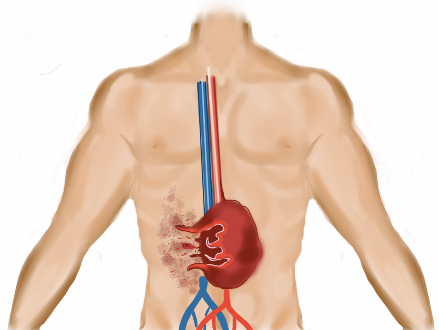
The incidence of abdominal aortic aneurysm (AAA) has been reported to be as high as 36.2 per 100,000 and is increasing.[1] Up to 62% of patients with ruptured aneurysms die before reaching the hospital, and the overall mortality rate after rupture may exceed 90%.[2] An AAA should be suspected in any patient > 60 years with complaints of abdominal or back pain, particularly if they have a history of hypertension or smoking. Ruptured AAA may present as undifferentiated hypotension and rapid diagnosis is essential.
Figure 3.1
Ruptured AAA
3.2 Indications/Anatomy
Indications for AAA scan:
- Abdominal Pain
- Back Pain
- Chest Pain
- Pulsatile Abdominal Mass
- Renal Colic
- Syncope
- Hypotension
- Weakness
- Neurologic Changes in the Extremities
Indications
The main impetus for the scan includes a clinical suspicion for AAA. Indications may include abdominal pain, back pain, chest pain, a pulsatile abdominal mass, “renal colic”, syncope, hypotension, weakness, or neurologic changes in the extremities. One high-risk group, in particular, includes older males with hypertension and a history of tobacco abuse.
Relevant Anatomy
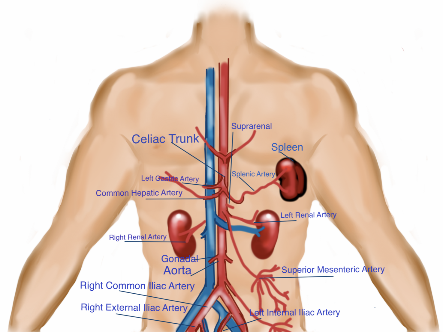
The abdominal aorta enters the abdomen through the diaphragm just inferior to the xiphoid process (T12) and bifurcates around the level of the umbilicus (L5). It has a predictable series of branches as pictured below (#Figure 3.2). The 1st branch off the abdominal aorta is the celiac trunk (#Movie 3.1).
Figure 3.2
Aortic Anatomy
Movie 3.1
Celiac Trunk
The celiac trunk divides into three vessels, although typically, only the hepatic artery and splenic artery are reliably visualized. The 2nd branch off the abdominal aorta is the superior mesenteric artery (SMA); typically, 1 cm caudal to the celiac trunk. The SMA runs parallel to the abdominal aorta and can be tracked caudally with the abdominal aorta. While the renal arteries may be visualized, the branches of the gonadal and inferior mesenteric arteries are not typically seen with ultrasound. The abdominal aorta ultimately bifurcates into the common iliac vessels around the level of the umbilicus.
3.3 Technique
Curvilinear probe is preferred.
Scan in transverse and longitudinal all the way through the bifurcation of the iliacs.
Use steady pressure to scan through bowel gas.
Aneurysms can be fusiform (more common) or saccular.
Probe Selection
Curvilinear probe 2-5 MHz (preferable) or phased array probe 1-5 MHz.
Technique
The general approach is to start with the probe indicator pointing to 9 o’clock, immediately caudal to the xiphoid process. This produces a transverse image of the abdominal aorta. You will need to scan from this mid-epigastric location caudally to the bifurcation of the aorta, visualizing the transverse high, mid, and low aorta (#Gallery 3.1, #Movie 3.2, #Movie 3.3).
Gallery 3.1
Technique
[[03_PosAAATxHigh.png]] | Transverse high probe placement
[[03_PosAAATxMid.png]] | Transverse mid probe placement
[[03_PosAAATxLow.png]] | Transverse low probe placement
Movie 3.2
Normal Scan of the Transverse Aorta
Movie 3.3
Iliac Bifurcation
Next, you will turn the probe towards the patient’s head at 12 o’clock (#Movie 3.4).
Movie 3.4
Normal Scan of the Proximal Aorta in Longitudinal
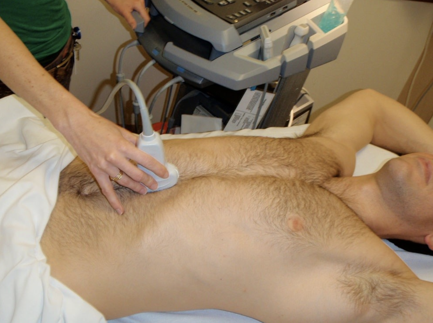
This produces a longitudinal view of the aorta. Again, you will scan from the mid-epigastrium to the bifurcation at the umbilicus (#Image 3.1 and #Movie 3.5).
Image 3.1
Longitudinal Probe Placement
Movie 3.5
Normal Longitudinal Scan of the Aorta to the Bifurcation
To adequately image the aorta, the first step is to identify the vertebral body. The vertebral body is horseshoe-shaped with an intense echogenic anterior surface and posterior shadowing (#Image 3.2).
The aorta is anterior to the vertebral body and slightly to the patient’s left (right side of the ultrasound image). The inferior vena cava (IVC) is also anterior to the vertebral body, and to the patient’s right.
Image 3.2
Normal Aorta with Spine Shadow
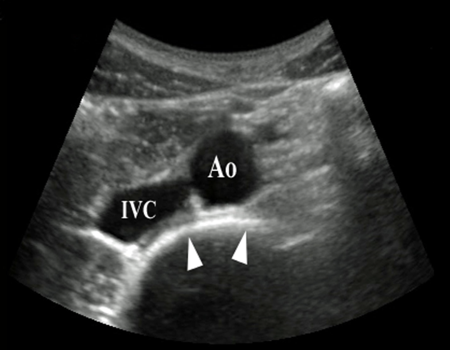
Scan caudally to the aortic bifurcation with methodical real-time visualization, without skipping any section of the aorta. As you scan cephalad to caudad (head to toe), identify the following: celiac trunk, (#Movie 3.1), SMA, and aortic bifurcation (#Gallery 3.2)
Gallery 3.2
Celiac Trunk, SMA, Aortic Bifurcation
[[03_ScanLongCeliacSMA.png]] | Transverse aorta with celiac trunk (seagull sign); Hep A = hepatic art, Spl A = splenic art,  * = aorta).
[[03_ScanPortalSMA.png]] | Ao = aorta, V = vert body, arrowhead = portal v, arrow = superior mesenteric artery.
[[03_ScanAoBifurcation.png]] | Level of the aortic bifurcation. I=iliac arteries, V = vertebral body.
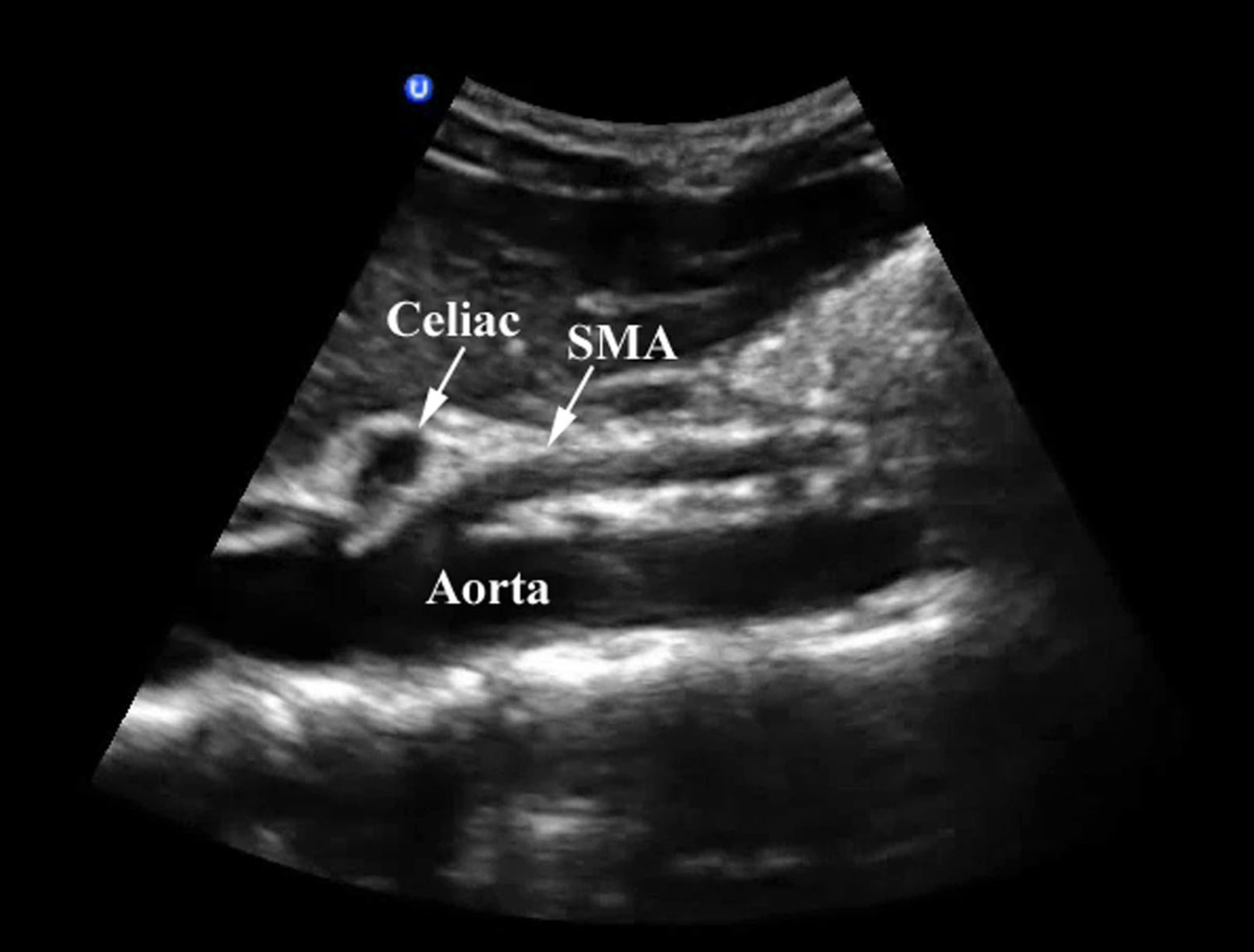
Turn the probe to the longitudinal plane (pointer to 12 o’clock) to obtain a longitudinal view of the aorta and the associated vessels (celiac and SMA) (#Image 3.3).
The same proximal branches (celiac and SMA) seen in transverse plane should be seen in longitudinal plane. Again, move cephalad to caudad as you scan the entire length of the vessel (#Movie 3.6).
Image 3.3
Proximal Aorta in Longitudinal
Movie 3.6
One Minute Ultrasound Transverse Aorta Scan Demo
#### Impediments & Solutions Ultrasound of the aorta can be quite difficult depending on bowel gas and body habitus. Imaging impediments due to bowel gas usually originate from the transverse colon, which sits in the epigastrium. If bowel is encountered, apply steady pressure (push down towards the patient’s back) with the probe. The bowel may be effectively compressed or undergo peristalsis. You may also try to jiggle the probe to move bowel aside. Another alternative is to fan through windows between loops of bowel. This is accomplished by establishing a good sonographic window, just cephalad to the area obscured by bowel, and tilting the probe toward the feet. Similarly, find a window just caudad to the obstructed area and angle the probe up toward the head. Obesity impediments may be addressed by asking the patient to lie completely flat. Have the patient flex their hips and knees to relax the abdominal muscles. Finally, lower the scanning frequency to increase penetration.Normal US Findings
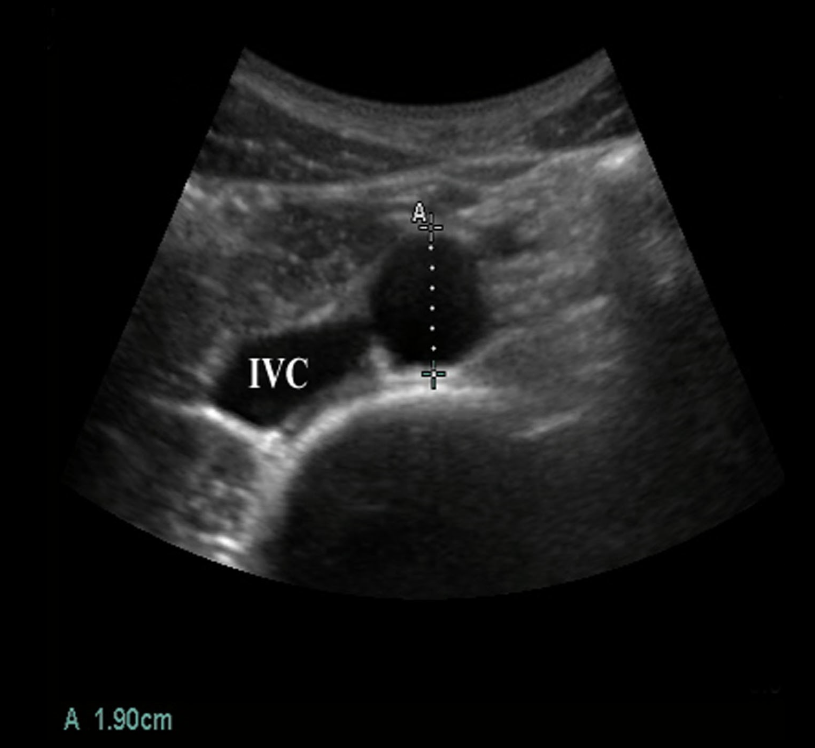
The normal aorta is < 3cm and tapers distally. Appropriate documentation will include a caliper measurement of the proximal, mid and distal aorta (#Image 3.4). Measurements should be taken in the transverse plane, to limit the tangential effect of underestimating diameter on a longitudinal image of the aorta. ALWAYS MEASURE OUTER WALL TO OUTER WALL. A mural thrombus or plaque in the lumen may underestimate the size of the AAA.
Image 3.4
Mid Aorta with Calipers with IVC Marked
Gallery 3.3
Fusiform and Saccular Aneurysms
[[03_ScanAAAShortLong.png]] | Fusiform and Saccular Aneurysms
[[03_AnatAAAFusiformCartoon.png]] | large segment of aorta affected
[[03_AnatAAASaccularCartoon.png]] | small segment of aorta baloons
[[03_ScanAAA.png]] | Fusiform and Saccular Aneurysms
[[03_ScanAAALong.png]] | Fusiform and Saccular Aneurysms
Abnormal US Findings
Any aortic measurement > 3cm is abnormal; any aorta that fails to taper appropriately as it moves caudally is considered abnormal as well. Of note, the majority of AAAs are INFRARENAL.3
There are two types of aneurysm: fusiform and saccular. (#Gallery 3.3). Fusiform aneurysms are much more common and involve dilation of the entire circumference of the affected segment of the aorta (#Gallery 3.4 and #Movie 3.7, #Movie 3.8, #Movie 3.9, #Movie 3.10, #Movie 3.11). A saccular aneurysm is an asymmetric outpouching (i.e. sac-like) of the aorta (#Gallery 3.3 and #Movie 3.12)
Gallery 3.4
Fusiform Aneurysms
[[03_ScanAortaDimension.png]] | Arrows noting AP diameter of AAA
[[03_ScanAortaCallipers.png]] | AAA in long with ( * ) noting lumen
[[03_ScanAortaDimensions2.png]] | 6.7cm AAA in longitudinal
[[03_ScanAAAFalseLumenDimensions.png]] | 7cm AAA; Lumen of aorta (arrows) and AAA as marked
[[03_ScanAAAFalseLumenDimensionsLong.png]] | Arrows indicate AAA, ( * *) demonstrates lumen.
Movie 3.7
3.5cm AAA in Transverse
Movie 3.8
7cm AAA in Transverse to Longitudinal
Movie 3.9
3.5cm AAA in Transverse
Movie 3.10
7cm AAA in Transverse
Movie 3.11
6cm AAA in Transverse to Longitudinal
Movie 3.12
Saccular Aneurysm
Of note, aneurysms may extend into the iliac arteries as well, so a comprehensive, methodical scan through the bifurcation is recommended (#Movie 3.13 and #Image 3.5).
Movie 3.13
7cm R Common Iliac Aneurysm
Image 3.5
7cm Iliac Aneurysm Marked
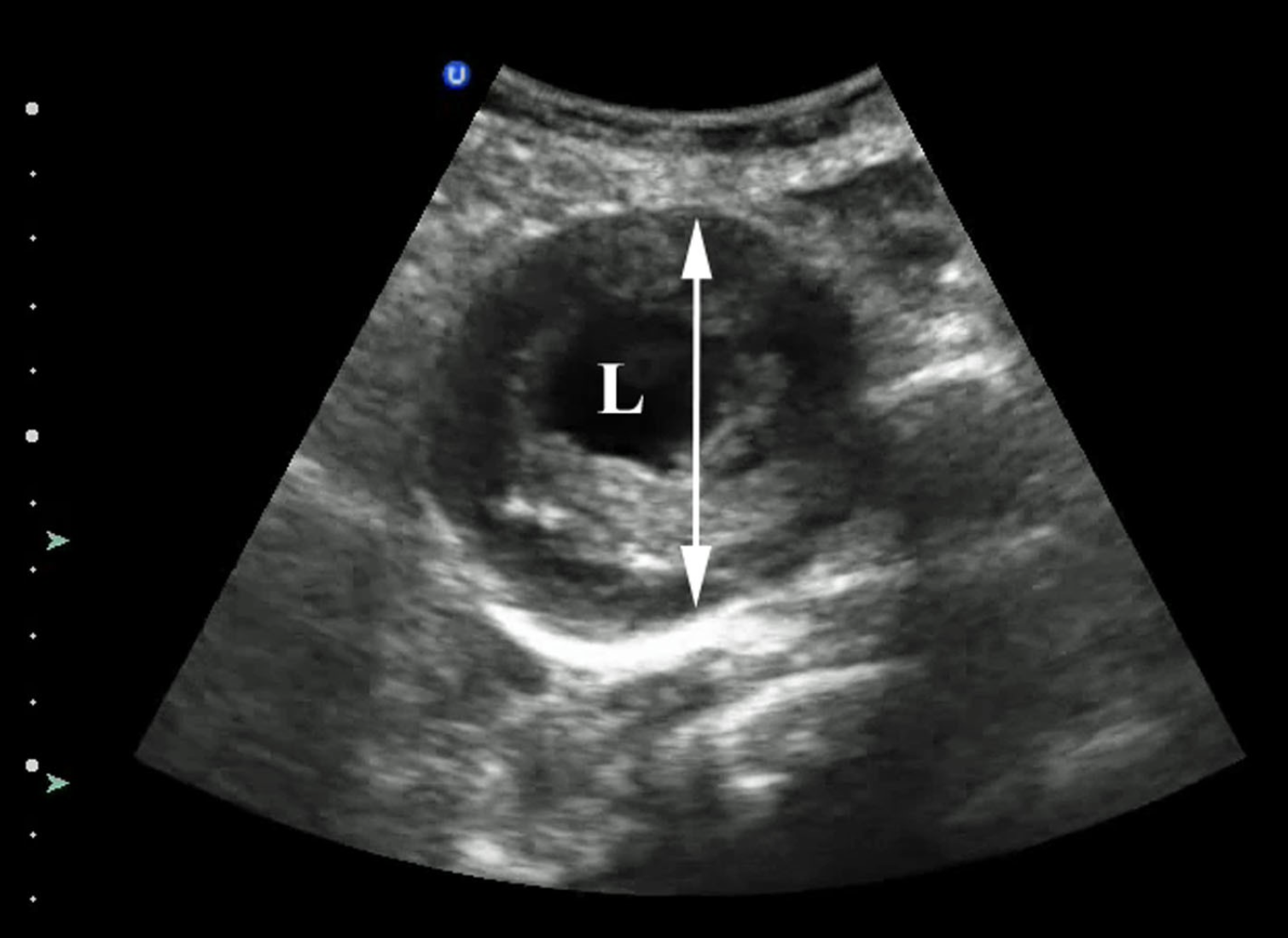
Image 3.6
Perihepatic Free Fluid (Arrowheads)
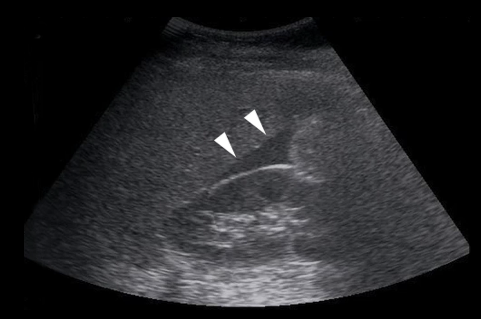
Emergency bedside ultrasound is highly sensitive for the presence of an AAA but has poor sensitivity for acute rupture.[3] Rupture of an AAA most commonly occurs into the retroperitoneal space and may tamponade before the patient becomes unstable. Unfortunately, ultrasound does not reliably identify retroperitoneal blood. See an ultrasound of a retroperitoneal bleed. An AAA rupture occasionally is intraperitoneal, with free fluid readily identified by ultrasound (#Image 3.6 and #Movie 3.14). This is a particularly ominous finding. An aortic dissection (floating intimal flap) may occasionally be found while performing an US to rule out AAA (#Gallery 3.5, #Movie 3.15 and #Movie 3.16).
Gallery 3.5
Aortic Dissection
[[03_ScanAorticFlapShort.png]] | Marked transverse aortic dissection (courtesy of Dr. Chris Moore, Yale School of Medicine)
[[03_ScanAorticFlapLong.png]] | Aortic dissection (longitudinal) marked by arrowheads (courtesy of Dr. Chris Moore, Yale School of Medicine)
[[03_ScanAorticFlapLong2.png]] | Arrowheads marking subtle aortic dissection.
[[03_ScanAorticFlap2.png]] | Arrowhead marking aortic dissection (courtesy of Dr. Chris Moore, Yale School of Medicine).
[[03_ScanAorticFlap3.png]] | ???
Movie 3.14
Intraperitoneal Free Fluid in Morrison's Pouch
Movie 3.15
Aortic Dissection in Longitudinal
Movie 3.16
Aortic Dissection Longitudinal to Transverse
3.4 Pearls and Pitfalls
- Be sure to measure outer wall to outer wall, as thrombus can cause underestimation of aneurysm size.
- Retroperitoneal rupture is hard to pick up on ultrasound.
- Always measure in transverse completely perpendicular to the vessel.
- Be careful to not mistake the IVC for the aorta.
- Dissection flaps are easily overlooked.
Thrombus within an AAA can be mistaken for the aortic wall, leading to an underestimation of the true AAA diameter. To avoid this error, adjust the gain so that aortic lumen is black. If possible, decrease the dynamic range to improve the contrast between vessel wall and lumen. Be sure to measure outer wall to outer wall.
Another pitfall is to assume an AAA is not ruptured in the absence of free intraperitoneal fluid. Retroperitoneal bleeding, which is a far more common site of rupture, is NOT reliably detected by ultrasound. Click here for a case study. Note: while ultrasound can reliably detect an AAA, it typically gives no information about rupture.
Saccular aneurysms can be easily missed unless a thorough scan is performed. Systematic, continuous scanning in both longitudinal and transverse planes is essential to prevent a false negative diagnosis.
An ectatic aorta may have irregular course (#Figure 3.3).
Figure 3.3
Ectatic Aorta
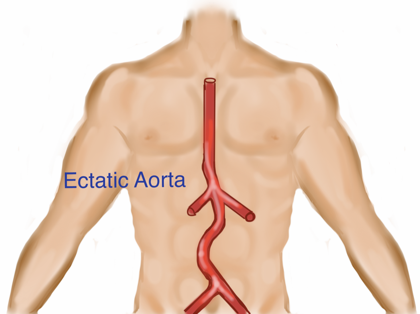
Dynamic scanning, while adjusting for changes in vessel angle, will allow for complete visualization.
Angled transverse cuts may exaggerate the true aortic diameter. Obtain measurements at 90° to the vessel. Off-axis (tangential) longitudinal cuts underestimate aortic diameter. Caliper measurements should be made in a transverse view only.
Inexperienced sonographers can mistake the IVC for the aorta, especially in long axis because both the aorta and IVC are pulsatile (#Image 3.7 and #Movie 3.17).
Image 3.7
Two Panels Comparing Sono Appearance of Aorta vs IVC
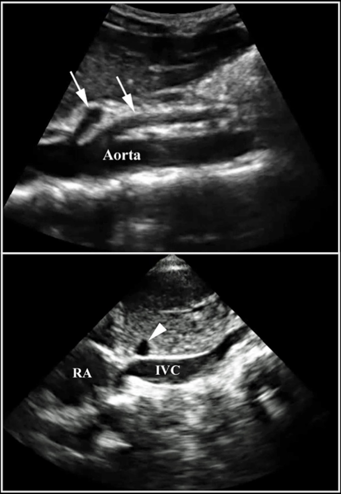
The aorta lies to the patient’s left of the IVC. Moreover, the aorta has anterior branches caudal to the liver, while the IVC does not. Finally, the aorta is typically more round, non-compressible, and has brighter, thicker walls than the IVC.
Small aneurysms (< 4.5cm) can rupture, although less frequently than a larger AAA.
If an AAA is identified by US, it still may not be the cause of the patient’s symptoms. Consider abdominal, renal or musculoskeletal pathology.
Dissection flaps are easily overlooked. If there is a high clinical suspicion for AAA and the ultrasound is equivocal, obtain a CT (with contrast if no renal impairment).
Movie 3.17
Comparison of Aorta to IVC
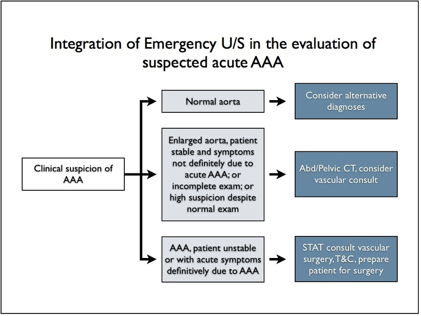
Figure 3.4
Flowchart of Aorta Ultrasound in the ED
3.5 References
Bickerstaff LK, Hollier LH, Van Peenen HJ, et al. Abdominal aortic aneurysms: the changing natural history. J Vasc Surg. 1984; 1(1):6-12. ↩︎
Ernst CB. Abdominal aortic aneurysm. N Eng J Med. 1993; 328(16):1167-1172. ↩︎
Kuhn M, Bonnin RL, Davey MJ, et al. Emergency department ultrasound scanning for abdominal aortic aneurysm: accessible, accurate and advantageous. Ann Emerg Med. 2000;36(3):219-223. ↩︎