Chapter 6. Renal

Chapter Authors & Contributors
- Ben Williams, MD
- Mike Mallin, MD, RDCS, RDMS
- Jonathan Wallace, MD, RDMS
- Rayna Sharma, BScKin
- Adam Guthrie, BKin, MMgmt
6.1 Introduction
Advantages of US over CT for renal:
- Shorter length of stay
- Lower cost
- Improved safety profile
Bedside ultrasound (US) can be utilized in the evaluation of patients with suspected kidney pathology to diagnosis causes of renal colic, renal failure, hematuria, and decreased urine output. In recent years, CT has replaced the physical exam and plain X-ray in the evaluation of these patients. However, ultrasound has many advantages over CT scanning including shorter length of stay, lower cost, and improved safety profiles.[1][2] Rosen and colleagues demonstrated a 147-minute reduction in length of stay when performing ultrasound in the place of CT for the evaluation of renal colic.1 Furthermore, recent literature on CT utilization has increased physician awareness of the ill effects associated with ionizing radiation exposure from CT scans.[3][4][5] For these reasons, there is growing interest in the use of US instead of CT for the evaluation of flank pain and suspected nephrolithiasis.
Renal US is becoming more commonly used and is considered a safe initial test in the evaluation of suspected nephrolithiasis and renal colic. In practice, US is commonly applied when the clinical suspicion for a kidney stone is high and the concern for another etiology of flank pain, such as an abdominal aortic aneurysm (AAA), is low. “Although stones in the kidney are easily visualized on US, when they pass into the ureter and cause pain they are often obscured by bowel gas and not readily seen. For this reason, the diagnosis of nephrolithiasis and renal colic on US is often made by secondary findings, such as hydronephrosis.[6]
Other diagnostic dilemmas in utilizing renal US to diagnose nephrolithiasis are the relative inaccuracy of predicting stone passage and the inability to evaluate for alternate causes of flank pain, such as AAA. Although CT may help to predict stone passage and evaluate the aorta, with a recurrent stone rate of 50% in most patients, performing CT scans on every patient with every presentation of flank pain would lead to astronomical radiation exposure.[7] This chapter will focus on the means by which a clinician can apply bedside US to evaluate patients with flank pain and suspected kidney stones, while attempting to minimize the risk of missed diagnoses and improper treatment of impassible stones.
6.2 Renal Anatomy
Two portions of the kidney:
- Renal parenchyma
- Collecting system
Hydronephrosis is dilation of the collecting system
The kidneys can be divided into two portions: the renal parenchyma and the collecting system. The renal parenchyma includes the cortex, which contains the filtration components of the glomerulus and the medulla. This is the area where the nephrons are located. The medulla contains the medullary pyramids, which are prominent hypoechoic structures seen on ultrasound, especially in the setting of hydronephrosis. The medullary pyramids contain the distal portions of the nephron and secrete urine into the minor calyces.
The collecting system begins with the minor calyces and ends at the hilum of the kidney where the ureter joins the renal pelvis. There are approximately 8-18 minor calyces that eventually coalesce into major calyces, which drain into the ureter [7:1] (#Figure 6.1 and #Image 6.1).
Figure 6.1
Renal Anatomy
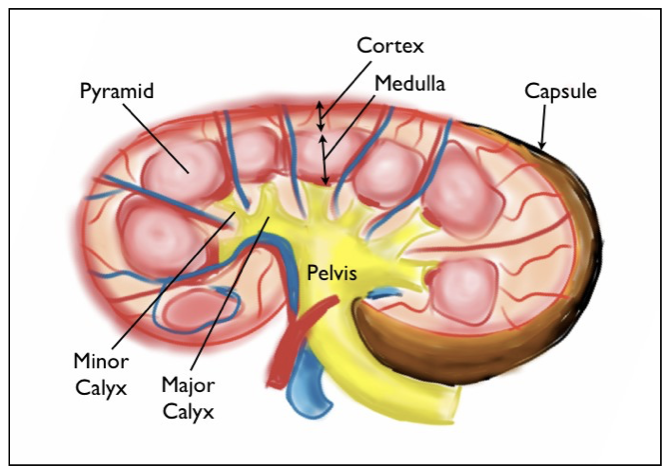
Image 6.1
Renal Anatomy
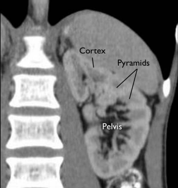
The renal cortex is a bright, or hyperechoic, structure with a ground-glass appearance that is located peripherally to the medulla. Typically, the renal cortex is just slightly darker than the liver or spleen, which are both readily available for comparison, as these organs are used as acoustic windows for visualization. The renal cortex is approximately 1-2 cm in width [8]([[#Movie 6.1]]).
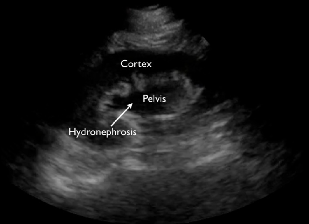
The renal pelvis is brighter, or more hyperechoic, than the cortex and is located centrally. The renal pyramids surround the pelvis and collecting system, which is often not visible in the normal kidney without hydronephrosis. Conversely, in the setting of obstruction and thus hydronephrosis, the collecting system is a dilated, fluid filled, and anechoic structure that dominates the central portion of the kidney (#Image 6.2).
Movie 6.1
Normal Kidney
Image 6.2
Hydronephrosis
Although kidney measurements are not typically performed at the bedside in clinical evaluation of flank pain, they can be helpful in the evaluation of other pathology such as causes of chronic kidney disease (CKD). Normal kidneys measure 9-12cm in length and 4-5cm in width with less than a 2cm variation when compared to the patient’s other kidney.[9] A large kidney suggests acute renal congestion from causes such as thrombosis, pyelonephritis, or acute renal failure.[10] Conversely, smaller kidneys suggest poor function and CKD.
The appearance and size of the renal cortex can also identify pathology. Normally, the thickness of the renal cortex is 1-2cm. A thin cortex can be seen in severe hydronephrosis and in CKD, whereas a large cortex can sometimes be seen with pyelonephritis. Finally, the difference in echogenicity between the cortex and the pelvis should be well demarcated in a normal kidney. However, chronic kidney disease can obscure this demarcation and cause the kidney to have uniform echogenicity.[11]
One Minute Renal Demonstration Video
6.3 Image Acquisition
- Curvilinear probe is preferred.
- Image in two planes.
- The left kidney is more posterior and superior.
- Image bladder in transverse and longitudinal orientation.
- Have a very low threshold for imaging the aorta in a patient with flank pain.
The curvilinear probe is ideal for imaging the kidneys. It uses a lower frequency and thus has improved penetration. This is necessary due to the retroperitoneal location of the kidneys, which must be imaged through acoustic windows such as the liver and the spleen. In situations where a curvilinear probe is not available, a phased array probe may be used as it also has a lower frequency (#Gallery 6.1).
Gallery 6.1
Probes
[[probeCurvilinear.jpg]] | Curvilinear transducer
[[Images/probePhased.jpg]] | Phased array transducer
Movie 6.2
Renal How-To Demo
The right kidney should be approached by placing the probe at the midaxillary line at the most inferior intercostal space. The probe marker should initially be oriented towards the patient’s head. Often a slight counterclockwise twist will allow the probe footprint to align between the ribs and obliterate any rib shadows in the image. From this position, the probe should be rocked from superior to inferior pole of the kidney and fanned from anterior to posterior to evaluate the entire kidney. Each kidney should be visualized completely in two planes. Thus, after this longitudinal view is obtained, the probe marker should be oriented anteriorly to create a transverse view of the kidney. Again, the probe should be fanned superior and inferior to visualize the entire kidney (#Image 6.3, #Image 6.4 and #Movie 6.3).
Image 6.3
Longitudinal Probe Placement
!700
Image 6.4
Transverse Probe Placement
!700
Movie 6.3
Normal Right Kidney
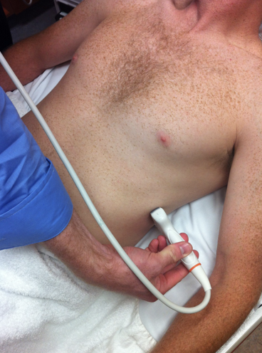
The left kidney uses the spleen as the acoustic window and is therefore more challenging. The probe should be initially placed over the posterior axillary line at the second most inferior intercostal space. Again, the probe will be oriented with the marker towards the patient’s head and then anteriorly. The left kidney should be visualized entirely in both longitudinal and transverse orientation (#Images 6.5, #Image 6.6, and #Movie 6.4).
Image 6.5
Longitudinal Left Kidney
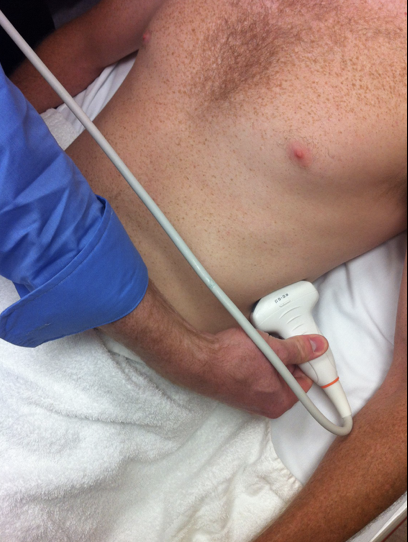
Image 6.6
Transverse Left Kidney
Movie 6.4
Normal Left Kidney
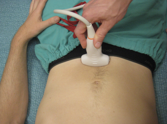
The bladder is another important structure that should be imaged when assessing for causes of hydronephrosis, renal colic and renal failure. To image the bladder, place the probe just superior to the pubis symphysis and direct the beam of the probe inferiorly, down into the pelvis. Acquire images in the longitudinal and transverse planes (#Image 6.7, #Image 6.8, #Movie 6.5 and #Movie 6.6).
The aorta should be imaged in virtually every patient with flank pain and suspected kidney stone. The exam is quick and highly sensitive (Chapter 3. Aorta).
Image 6.7
Longitudinal Bladder
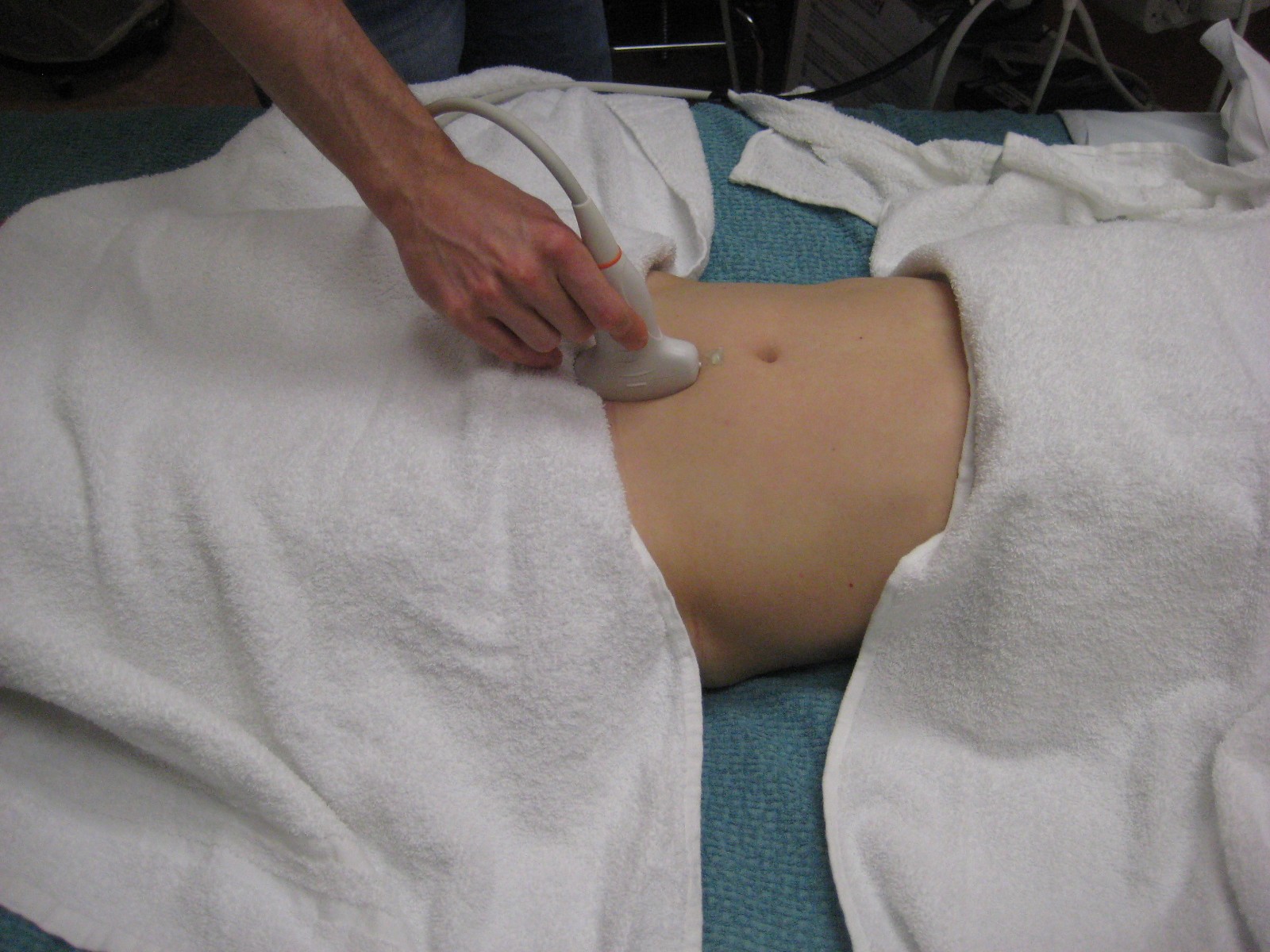
Image 6.8
Transverse Bladder
Movie 6.5
Longitudinal Bladder
Movie 6.6
Transverse Bladder
6.4 Pathology
- 5-15% of Americans will have a kidney stone at some point in their life.
- The evolution of renal colic is focused on secondary signs such as hydronephrosis
- Severe hydronephrosis is characterized by cortical thinning
- Avoiding CT scans on patients with no or mild hydronephrosis may decrease CT utilization by 73%
- Do not confuse cysts with hydronephrosis
Kidney Stones
A common indication for bedside renal US is in the evaluation of flank pain and suspected renal colic. It is estimated that 5-15% of Americans will have a kidney stone at some point in their lifetime.[5:1][12] Symptoms of renal colic include: sudden unilateral flank pain, inability to achieve a comfortable position, radiation of pain to the groin, hematuria/dysuria, and nausea and vomiting.
The ultrasound evaluation of renal colic is directed more towards secondary findings, such as hydronephrosis due to ureteral obstruction. However, kidney stones can sometimes be visualized in the collecting system or in the proximal ureter. Kidney stones appear as hyperechoic structures that vary in size from 1mm-10mm and cast a prominent shadow. Unfortunately, US is not reliable at identifying the actual stone. The sensitivity of ultrasound to identify renal stones in the kidney is about 60-67%.[13][14] The sensitivity is even worse when diagnosing ureteral stones. In a study by Smith et al,[6:1] ultrasound was reported to be only 19% sensitive for finding ureteral stones.
Due to the relative insensitivity of US diagnosis of actual stones, the operator should instead depend on the presence of unilateral hydronephrosis on the side of pain and a high clinical suspicion for obstructing ureteral stone as more conclusive proof than visualization of the stone itself. Luckily, bedside US has proven to be highly sensitive for the diagnosis of hydronephrosis. The sensitivity of identification of hydronephrosis is 71-97%.[15][16]
Figure 6.2
Hydronephrosis
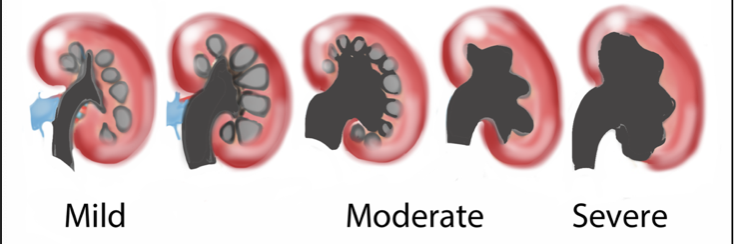
Hydronephrosis is a descriptive finding, not etiology, caused by obstruction of the ureter, bladder, or urethra, and thus urine backup into the renal pelvis. Acutely, this can cause the calyceal system to dilate with urine, which appears anechoic on ultrasound. The severity of hydronephrosis occurs along a spectrum described as mild, moderate or severe, which can be delineated by the renal structures that are affected[17] (#Figure 6.2). Hydronephrosis may appear to be absent in the setting of ureteral obstruction and volume depletion due to the relative lack of backup of urine into the collecting system. For this reason, many physicians provide a patient with a fluid bolus prior to bedside imaging to improve sensitivity.[18] Mild hydronephrosis is characterized by enlargement of the calices with preservation of the renal papillae (#Movie 6.7 and #Movie 6.8).[17:1]
Movie 6.7
Mild Hydronephrosis
Movie 6.8
Mild Hydronephrosis
Moderate hydronephrosis is characterized by rounding/blunting of calices with obliteration of the papillae, and often described as having a bear-claw appearance (#Movie 6.9).[10:1][17:2]
Movie 6.9
Moderate Hydronephrosis
Severe hydronephrosis is the most dramatic and is characterized by caliceal ballooning with cortical thinning (#Movie 6.10, #Movie 6.11 and #Movie 6.12).[10:2]
Movie 6.10
Severe Hydronephrosis
Movie 6.11
Severe Hydronephrosis
Movie 6.12
Severe Hydronephrosis
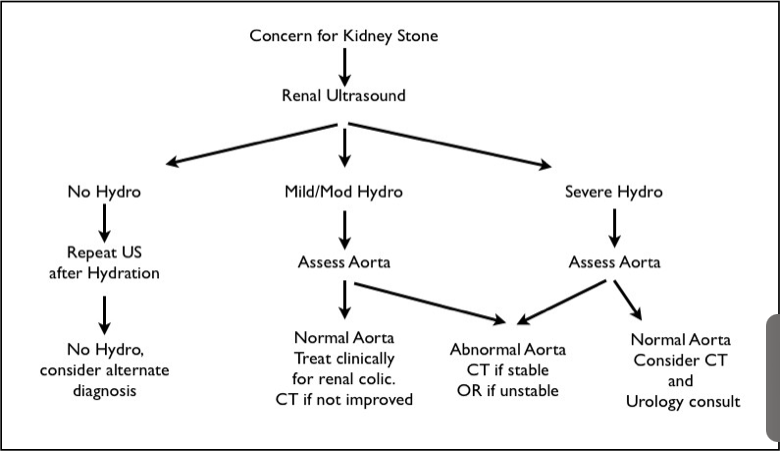
Unfortunately, there is no correlation between the degree of hydronephrosis and renal function, or extent of ureter obstruction.[10:3] However, Goertz and Lotterman reported that the degree of hydronephrosis on ultrasound was associated with a proportional increase in stone size and thus the likelihood of stone passage.[17:3] Typically, small stones <5mm are expected to pass spontaneously and large stones >10mm require surgical intervention for passage. Goertz and Lotterman suggested that patients be separated into less severe hydronephrosis (none or mild) and more severe hydronephrosis (moderate to severe). They noted that patients with more severe hydronephrosis frequently had large stones (>10mm) that were unlikely to spontaneously pass. Conversely, they also found that patients with less severe hydronephrosis could avoid CT scans during their workup, as there were no patients in this group with nephrolithiasis >10mm. Avoiding CT scans on patients with no or mild hydronephrosis thus would decrease CT utilization by 73% and only miss 9% of stones >5mm. #Figure 6.3 depicts this approach.
Figure 6.3
Approach to Patients with Symptoms Concerning for Kidney Stone
Renal Cysts
Renal cysts are the most common renal pathology found on ultrasound.[9:1] They can be asymptomatic or a cause of flank pain. There are two types of renal cysts, simple or complex.
Simple cysts must meet four criteria:
- Uniform, smooth oval shape.
- Anechoic center without internal echoes or septation.
- Well-demarcated border separating it from the surrounding renal parenchyma.
- Posterior acoustic enhancement, as seen with other fluid filled structures.
Failure to meet all four of these criteria would constitute a complex cyst. If there is a complex cyst, consider ordering a CT or MRI, as ultrasound cannot accurately characterize complex cystic masses.[^19]
In #Movie 6.13 demonstrating cortical cysts, note how the anechoic fluid collections are present in the renal parenchyma, not the pelvis. This differentiates renal cysts from hydronephrosis.
Movie 6.13
Cortical Cysts
Renal cysts can be problematic, as they can be mistaken for hydronephrosis. It is important when evaluating for hydronephrosis to image the kidney completely and convince yourself that the fluid filled structure is within the collecting system, as would be seen with hydronephrosis, and not within the cortex, as would be seen with a cyst.
Conclusion
In conclusion, renal ultrasound can be used to help diagnosis the etiology of flank pain. It has the benefit of not subjecting the patient to ionizing radiation, but should be appropriately applied. The aorta should virtually always be visualized to ensure the patient’s flank pain is not due to aortic pathology. In most cases of flank pain, patients with a history of kidney stones, unilateral mild-moderate hydronephrosis and a normal aorta can be treated clinically with appropriate follow-up. A multicenter trial, the STONE (Study of Tomography Of Nephrolithiasis Evaluation) trial, is currently investigating the diagnostic accuracy and safety of an ultrasound only approach.
6.5 REFERENCES
Rosen CL, Brown DFM, Sagarin MJ, et al. Ultrasonography by emergency physicians in patients with suspected ureteral colic. J Emerg Med. 1998;16(6):865-870. ↩︎
Pearle MSP, Calhoun EA, Curhan GC, et al. Urologic diseases in America project: urolithiasis. J Urol. 2005;173:848-57. ↩︎
Smith-Bindman R, Lipson J, Marcus R, et al. Radiation dose associated with common computed tomography examinations and the associated lifetime attributable risk of cancer. Arch Intern Med. 2009;169(22):2078-2086. ↩︎
Committee to Assess Health Risks from Exposure to Low Levels of Ionizing Radiation, National Research Council. Health Risks from Exposure to Low Levels of Ionizing Radiation: BEIR VII Phase 2. Washington, DC: The National Academies Press; 2006. ↩︎
Einstein AJ, Henzlova MJ, Rajagopalan S. Estimating risk of cancer associated with radiation exposure from 64- slice computed tomography coronary angiography. JAMA. 2007;298(3):317-323 ↩︎ ↩︎
Smith RC, Verga M, McCarthy S, et al. Diagnosis of acute flank pain: value of unenhanced helical CT. AJR Am J Roentgenol. 1996;166(1):97–101. ↩︎ ↩︎
Swadron S, Mandavia DP. Renal ultrasound. In: Ma OJ, Mateer JR, editors. Emergency Ultrasound. New York: McGraw Hill Professional; 2002:197–220. ↩︎ ↩︎
Bluth E, Benson C, Ralls P, et al. Ultrasound: A Practical Approach to Clinical Problems. 2nd edition. New York, NY. 2008:84-111. ↩︎
Brant WE. Ultrasound: The Core Curriculum. Philadelphia, PA: Lippincott Williams & Wilkins, 2001:103-141. ↩︎ ↩︎
Noble V. Renal ultrasound. Emerg Med Clin N Am. 2004:22:641–65. ↩︎ ↩︎ ↩︎ ↩︎
Coleman BG. Ultrasonography of the upper genitourinary tract. UrolClin N Am. 1985;12:633–44. ↩︎
Teichman JM. Clinical practice. Acute renal colic from ureteral calculus. N Engl J Med. 2004;350:684-93. ↩︎
Sinclair D, Wilson S, Toi A, et al. The evaluation of suspected renal colic: ultrasound scan versus excretory urography. Ann Emerg Med. 1989;18:556–9. ↩︎
Lanoix R, Leak LV, Gaeta T, et al. A preliminary evaluation of emergency ultrasound in the setting of an emergency medicine training program. Am J Emerg Med. 2000;18(1):41–5. ↩︎
Gaspari RJ, Horst K. Emergency ultrasound and urinalysis in the evaluation of flank pain. Acad Emerg Med. 2005;12(12):1180-4. ↩︎
Goertz JK, Lotterman S. Can the degree of hydronephrosis on ultrasound predict kidney stone size? Am J Emerg Med. 2010;28(7):813-816. ↩︎
Middleton W, Kurtz A, Hertzberg B. Ultrasound: The Requisites. 2nd edition. Philadelphia, PA: Mosby; 2004:103-190. ↩︎ ↩︎ ↩︎ ↩︎
Israel GM, Bosniak MA. How I do it: Evaluating renal masses. Radiol. 2005;236: 441-450. ↩︎